您当前的位置: 首页 > 学校概况

学校简介:
学校教员: 人,学生数量:人, 校友数量: 人,子机构数量:个, 相关机构: 个,受资助项目:项, 文章数: 篇,专利数:项,
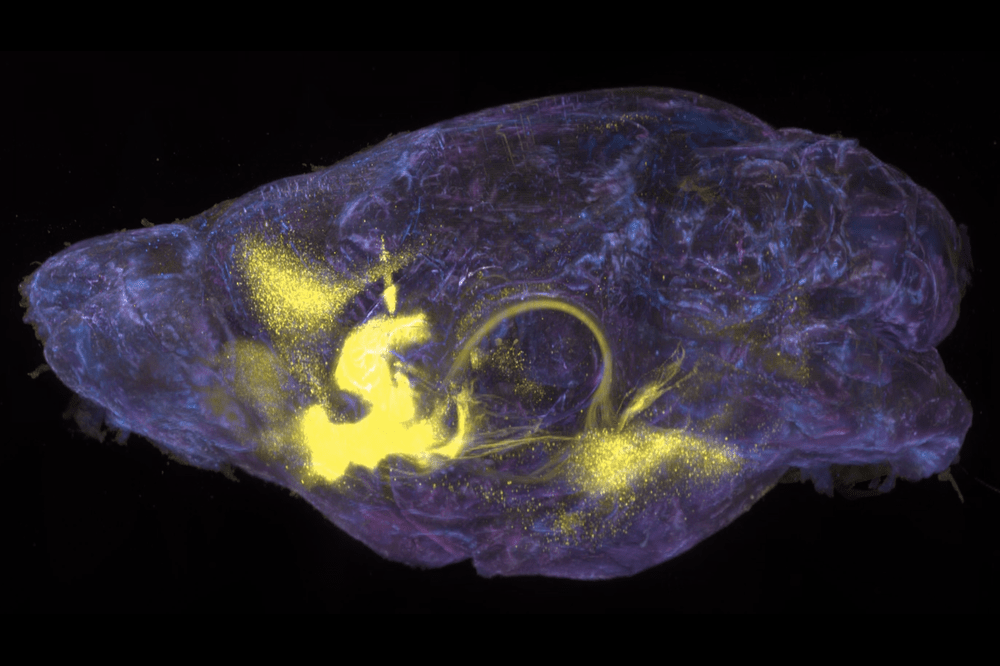
Drugs like morphine and cocaine fundamentally warp the brain’s reward system—creating the urge to use while,simultaneously,throwing natural urges to eat and drink off-kilter.Now,scientists from The Rockefeller University and Mount Sinai have identified,for the first time,a common reward pathway that may serve as ahub for rearranging such fundamental priorities.The findings,published in Science,shed light on neural processing of diverse classes of rewards in mice,with potential implications for understanding substance use disorders in humans.“We’ve known for decades that natural rewards,like food,and drugs can activate the same brain region,”says Rockefeller’s Jeffrey F.Friedman.“But what we’ve just learned is that they impact neural activity in strikingly different ways.One of the big takeways here is that addictive drugs have pathologic effects on these neural pathways,that’s distinct from,say,the physiologic response to eating ameal when you are hungry or drinking aglass of water when you are thirsty.”Homing in on the reward circuit Nestled in the forebrain,the nucleus accumbens(NAc)is involved in processing rewards from and desire for food,sex,social interaction—and addictive substances.The NAc,in close collaboration with the pleasure and mood modulating neurotransmitters dopamine and serotonin,influences decision-making by integrating motivation,reinforcement,and pleasure,essentially encouraging animals to repeatedly pursue activities that routinely feel good.“The NAc is akey node where the underlying dopaminoceptive neurons direct and refine animals’behaviors towards their goals,”says Bowen Tan,a graduate student in Friedman’s lab.“What we hadn’t been able to understand is how repeated exposure to drugs corrupts these neurons,resulting in escalated drug-seeking behaviors and ashift away from healthy goals.”To answer that question,Friedman and Tan teamed up with Mount Sinai’s Eric J.Nestler,a psychiatrist and expert on the molecular neurobiology of drug addiction and depression.Together they turned to Rockefeller’s Alipasha Vaziri to overcome technical limitations that have hampered past work in the field.Brain imaging techniques developed in Vaziri’s lab are among the only tools capable of capturing the majority of the mouse cortex in real-time with high resolution.But in this case,the researchers also needed the capability to record neurons at large tissue depths,in order to image the neural activity at single-cell resolution in the NAc.“Advancing our understanding of the intricately connected network within the brain demands the innovation of cutting-edge imaging technologies that can capture neuronal activity across distant brain regions,but also that of those at deeper regions,”Vaziri says. 查看详细>>
来源:洛克菲勒大学 点击量: 0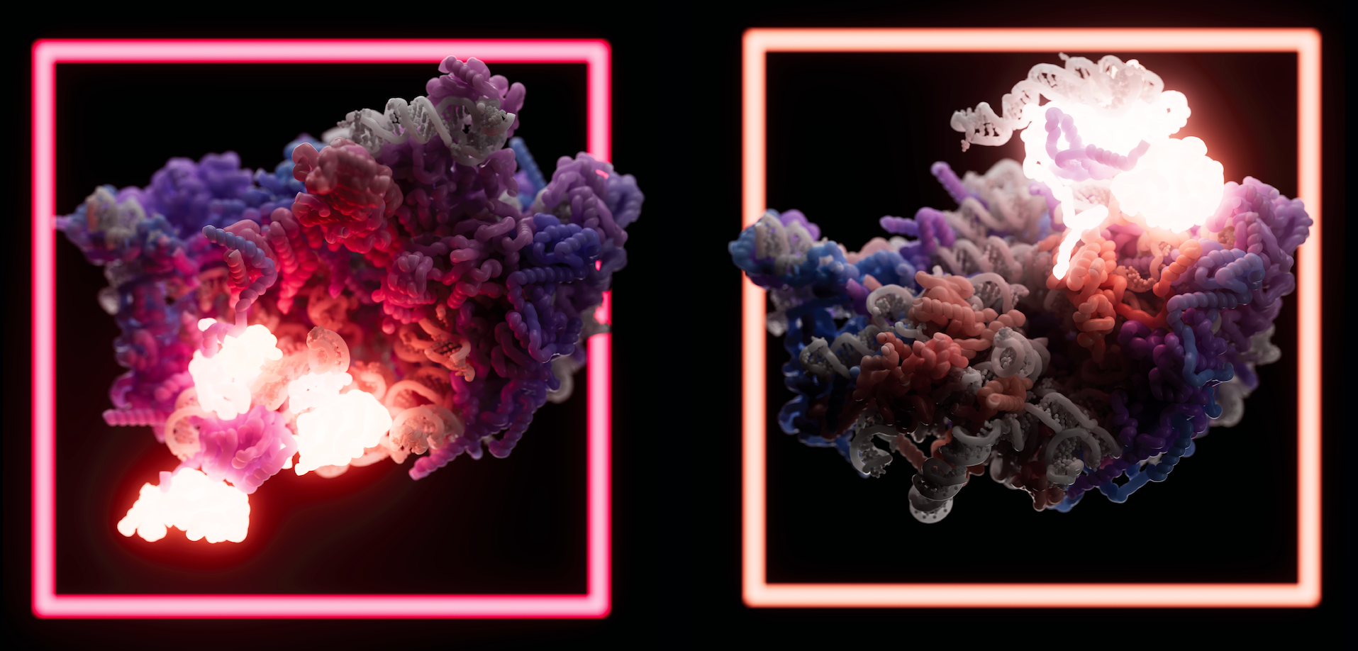
Life runs on ribosomes.Every cell on earth needs ribosomes to translate genetic information into all the proteins needed for the organism to function—and to in turn make more ribosomes.But scientists still lack aclear understanding of how these essential nanomachines are assembled.Now,new high-resolution images of the large ribosomal subunit are shedding light on how arguably nature’s most fundamental molecule coalesces in human cells.The findings,published in Science,bring us one step closer to acomplete picture of ribosome assembly.“We now have apretty good idea of how the large ribosomal subunit is assembled in humans,”says Rockefeller’s Sebastian Klinge.“We still have quite afew gaps in our understanding,but we certainly now have amuch better idea than we had before.”Solving the large subunit Ribosomes were first discovered at Rockefeller almost 70 years ago.Scientists have since determined that they are composed of two subunits:a small subunit,known as 40S,which is responsible for decoding messenger RNA,and alarge subunit(60S),which attaches protein pieces together.But those were the very broadest strokes.The precise steps by which these complex molecules are assembled into their mature form has long remained amystery.Klinge’s approach to this larger problem has long focused on figuring out how ribosomes form in the first place.To that end,Klinge’s lab was among the first to use cryo-electron microscopy to capture footage of anonbacterial ribosome assembling towards its final shape,and the lab has since taken an even more granular approach—painstakingly stringing snapshots of maturing ribosomes together,to understand how these molecules get from one point in their assembly to the next.In recent years,Klinge and other scientists around the world have identified and characterized more than 200 ribosome assembly factors that influence the modification,processing,and folding of ribosomes.For the current study,Klinge and colleagues focused on the human large ribosomal subunit(60S).The team already knew,from studies in yeast,that the large subunit’s formation involves two precursors(a 5S rRNA and 32S pre-rRNA)snapping together,but“we wanted to know all of the events that need to happen for this to occur,”says Arnaud Vanden Broeck,a postdoctoral researcher in Klinge’s lab.“We wanted to explain how the large subunit is assembled and processed in human cells.”Vanden Broeck and Klinge combined new techniques involving amashup of genome editing and biochemistry,to capture high-resolution cryo-EM structures of 24 human large ribosomal subunit assembly intermediates as they were maturing.The resulting images show how assembly factors,various proteins and enzymes,interact with RNA elements to drive the formation and maturation of the 60S.Together,the findings represent anear-complete picture of how the human large subunit assembles.“For sixty years we had almost nothing on the intermediates that form the human 60S—it was all but invisible to us—and now we’ve jumped from nothing to pretty good coverage,”Vanden Broeck says,while admitting that some of the rarest and most transient steps on the road to the mature 60S may have evaded the team,and fallen through the cracks.“We still have alot of work to do.”Nonetheless,key findings from the study could already begin informing related fields of inquiry.Among the intermediary steps discovered,for instance,are signaling pathways that suggest alink between ribosome assembly and cellular metabolism—suggesting that acomplete understanding of ribosomes may well require close collaboration with experts in cell metabolism.And the granular look at the steps of ribosome formation provided by the study may provide important context for scientists studying diseases linked to ribosome mutations.For now,however,Klinge and Vanden Broeck are content to marvel at the substantial leap forward.“It’s not guesswork anymore,”Klinge says.“We can now see,in detail,what’s going on when the large subunit assembles.It’s humbling to realize we’re finally able to see what makes ribosomes and drives protein formation in all of our own cells.” 查看详细>>
来源:洛克菲勒大学 点击量: 0
Rockefeller has topped asurvey that weighs what percentage of an institution’s scientific publications are frequently cited by other researchers.According to the CWTS Leiden Ranking of over 1,400 universities from 72 countries,Rockefeller has the highest percentage of most frequently cited scientific publications as aproportion of the total number of its publications.This impact ranking underscores how the findings of Rockefeller scientists are seen as significant and relevant to others in the scientific community.Each year,the ranking is conducted by the Netherlands-based Center for Science and Technology Studies of Leiden University.While Rockefeller was not included in the rankings due to achange in eligibility criteria in 2019,every other year since 2016,Rockefeller topped the Leiden ranking in the same categories.Rockefeller has also repeatedly excelled in similar reports such as U-Multirank.In the 2023 Leiden ranking,which examined the time period of 2018 to 2021,31%percent of Rockefeller publications were among the top 10 percent most frequently cited of all scientific publications.In second place,MIT,had 23.3 percent;and Princeton,Caltech,and Stanford University were close behind.When measuring the proportion of publications in the top one percent,Rockefeller also took first place with 5percent of university publications in the top one percent.MIT,Princeton,Caltech,and Stanford University were also in the top five.The Leiden Ranking’s methodology accounts for differences between scientific fields in citation and collaboration practices.Institutional publication data was collected from the Web of Science database. 查看详细>>
来源:洛克菲勒大学 点击量: 2
Most people get infected with human papilloma virus at some point in their lives but,thanks to arobust immune response,are none the wiser.Unless giant horn-like growths sprout from their skin.An exceedingly rare feature of HPV known as tree-man syndrome,this condition is afar cry from the skin and genital warts that often accompany symptomatic disease.How could one virus provoke such asevere reaction in aselect few,but leave most people symptom-free?In anew study published in Cell,Rockefeller’s Jean-Laurent Casanova answers this question by examining the genetics of an otherwise healthy patient who contracted asevere case of tree-man syndrome and several family members who exhibited milder reactions to HPV.Casanova’s team identified amutation that affects one’s reaction to HPV by decreasing the production of CD28,a molecule within the immune system that plays an important role in activating pathogen-fighting Tcells.Given the purported importance of CD28 to the immune system,the scientists were surprised that this CD28-deficient individual was healthy prior to contracting tree-man syndrome.“CD28 is thought of as apillar of Tcell immunity,”says Casanova.“The fact that this patient was otherwise healthy suggests that CD28 is largely redundant in human health.Something else is able to step up to provide protection against other infections.”The findings play into the larger body of Casanova’s work,which continues to demonstrate that the severity of influenza,tuberculosis,COVID-19,and other diseases,is not solely dependent on the pathogen itself,but on genetics of the host,too. 查看详细>>
来源:洛克菲勒大学 点击量: 18
Perpetually hungry,worms are strategic when it comes to searching for food.The microscopic roundworm Caenorhabditis elegans,or C.elegans,is known to spend up to 20 minutes seeking out snacks in its immediate surroundings before endeavoring to look elsewhere.Now,Rockefeller scientists have identified circuits in the C.elegans brain that underlie this behavior.In anew study,published in Neuron,the researchers describe neural mechanisms responsible for local search,showing that this response can be triggered by either smell-or touch-related cues.Looking for grub in all the wrong places C.elegans feed on bacteria.And when their environment is teeming with edible microbes,they needn’t move much to remain sated.But when food availability decreases,the worms start wiggling around abit.First,they extensively search the area in which they last encountered sustenance,circling asmall region for 10 to 20 minutes.Eventually,however,the worms widen their search perimeter—a strategy that resembles,and is possibly related to,some aspects of human behavior.“If you lose your keys,you first search locally:in your wallet,then in your coat,and other nearby places.And you do this repeatedly,”says Cori Bargmann,the Torsten N.Wiesel Professor.“But if that doesn’t work,at some point you begin searching abroader area.”The switch from local to global search has been observed in hungry insects,reptiles,fish,and mammals,suggesting that it may represent aconserved foraging strategy.With auniquely small and well-characterized nervous system,C.elegans provided Bargmann and her colleagues with handy tools to study the basic brain mechanisms driving this behavior.Munching on microbes To identify cellular components involved in local search,biomedical fellow Alejandro López-Cruz first analyzed the genes of C.elegans mutants that failed to produce this behavior—worms that skipped the whole“local”phase and proceeded directly to global search.These animals,he discovered,had adefective form of aprotein known as MGL-1,a receptor for the neurochemical glutamate.Additional experiments showed that MGL-1 receptors are found on two C.elegans neurons involved in detecting food:one called AIA,which receives input from chemosensory cells sensitive to food odors;and another called ADE,which is connected to mechanosensory cells responsive to food texture.Mechanosensory neurons and ADE,the researchers found,are not physically connected to chemosensory neurons and AIA—suggesting that smell and touch responses function independently.Supporting these findings,the researchers showed that animals using only one sensory system can effectively detect and react to food removal.“If you knock out glutamate from chemosensory and mechanosensory cells then you lose the behavior.But if you knock out only one,the animals respond normally,”says López-Cruz.“This tells us that there are two parallel and redundant pathways for detecting food.”“That makes sense,”adds Bargmann,“because food is probably too important to survival to represent it in just asingle form.”Memorable meals Investigating the longevity of local search,the researchers found that when mechanosensory or chemosensory neurons are stimulated,they act on MGL-1 receptors to modify the activity of ADE or AIA cells for minutes at atime.This cellular modification,in turn,triggers other neurons responsible for local search.In short:When food goes missing,worm brain activity is altered for an extended period of time,yielding an extended search period.This process,says López-Cruz,seems to represent the formation of amemory related to changes in food availability.“When we take the worm’s food away,we are essentially giving it afood-removal stimulus.The behavioral change that follows outlasts that stimulus by fifteen minutes,which is consistent with short-term memory,”he says.The switch from local to global search,says Bargmann,seems to reflect the process of forgetting.“A worm’s memory defines the period of local search,”she says.“And when it forgets,the animal moves on to longer-range exploration.” 查看详细>>
来源:洛克菲勒大学 点击量: 266
Infections caused by MRSA bacteria(pictured)can lead to serious health complications.Lysins kill the microbes by destroying the bacterium’s cell wall(green).Humans are in aconstant arms race with infectious bacteria.To kill these disease microbes,we develop powerful antibiotics;and in turn,the bacteria develop resistance against these drugs.So we enhance our antibiotics,and the bacteria enhance themselves accordingly—resulting in so-called superbugs.Increasingly,medications fail to eliminate these highly adapted bacteria,leaving our bodies dangerously defenseless.In light of this crisis,for almost 20 years Rockefeller’s Vincent A.Fischetti has been developing anovel form of antimicrobial ammunition known as lysins.Now,these bacteria-killing enzymes have been studied in aphase II human clinical trial,becoming the first antibiotic alternatives to achieve successful outcomes in this stage of clinical development.Natural born killer Some viruses are very good at killing bacteria.Known as bacteriophages,or simply phages,these viruses infect amicrobe,replicate inside of it,and then produce lysin enzymes,which cleave the bacterium’s cell wall.As aresult,progeny phages are released from within the bacterium,and the bacterium itself perishes.In nature,this kind of assault is commonplace:bacteriophages inhabit everything from oceans and soil to human bodies,helpfully regulating microbe populations wherever they go.In fact,every 48 hours half the bacteria on earth are killed by phages,making lysins the most widespread bacteria-killing agents on Earth.In the lab,lysins can be used as atool to break down and study the cell walls of bacteria—which is exactly what Fischetti was doing at Rockefeller about two decades ago.Simultaneously,his lab was also working on avaccine for streptococcus infections,and the broader research community was becoming increasingly worried about antibiotic-resistant infections.This confluence of events led Fischetti to abreakthrough.“Since Iwas working with lysins,I knew they killed bacteria instantly.My lab happened to have animals that were orally colonized with streptococci for my vaccine studies,”he recalls.“So,I thought,let me just give these colonized mice some lysin and see what happens to the streptococci.”The effect was dramatic:an hour after getting the drug,the animals were decolonized of their streptococci.The subsequent publication of this finding was the first to report the therapeutic use of phage lysins.Compelled by this result,Fischetti and later other scientists began developing lysins against several types of drug-resistant bacteria,many of which successfully cured infections in awide range of animal models.Until recently,however,no one had tested whether this type of therapy was safe and effective in humans. 查看详细>>
来源:洛克菲勒大学 点击量: 0